Learn why dentists may x-ray your teeth
When your dentist examines your mouth, they are looking for changes and potential problems which may need treatment. Regular appointments help to diagnose issues before they become serious. However, there’s a limit to how much can be seen with the naked eye, no matter how thorough the examination. This is why x-rays are so valuable, they help to see between and inside the teeth where tooth decay (holes) is hidden and commonly occurs.
Although the enamel on the tooth surface may appear intact, an x-ray can help to pick up signs of decay underneath. X-rays also help to detect problems with the roots of the teeth, changes in the bone, unerupted teeth such as wisdom teeth, infections and if there are issues with the jaw and facial bones. X-rays also assist with planning for dental care, including fillings, implants, tooth extraction and orthodontics.
Are dental x-rays safe?
Absolutely. Modern digital x-rays use very small amounts of radiation to create an image of the teeth and bones of the mouth and are safe for adults and children.
Radiation is measured in units called ‘Micro-Sieverts’. Walking around each day exposes us natural background radiation of 10 micro-sieverts whilst eating a banana is equivalent to 0.1 micro-sieverts. To put this into perspective, a typical single dental x-rays is equivalent to the natural radiation of eating two bananas.
If you are pregnant or breastfeeding, let your dentist know before you have any dental examination, including x-rays. According to health experts, for women who have not had a recent dental check and where there are benefits in having an x-ray, there is no need to defer having one. 1
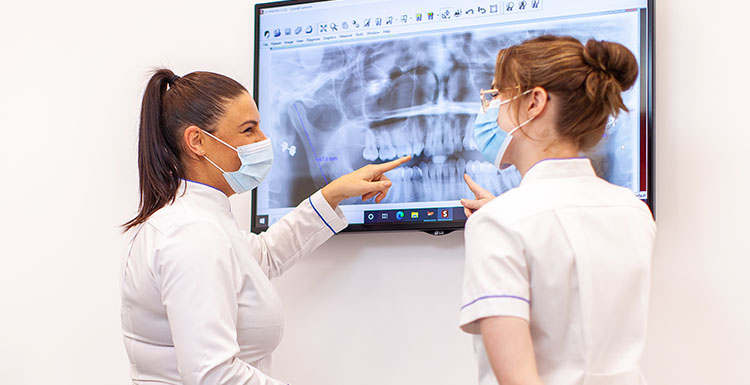
When your Dentist look at x-rays, they are looking for subtle changes in how light or dark an area on the x-ray appears to the surrounding area. This tells them if there has been any change in the mineral structure of the teeth and bone.
What type of dental x-ray would I need?
The type of x-ray you have will be based on the information your dentist needs.
- The most common type is what’s known as ‘Bite wing’ x-rays, which are generally taken at your first visit and then at intervals of between 12-24 months depending on your tooth decay risk factors.
- Periapical x-rays help to view the entire tooth, including the roots and surrounding bone. These types of x-rays are useful for helping to diagnose infections, bone levels, tooth aches or root structures and are required before a tooth is extracted.
- Panoramic x-rays, also called a ‘full mouth x-ray’ or OPG, help to visualise the entire mouth, sinuses, bones and jaw and to assess the stages of tooth development in children. They’re also useful to view the wisdom teeth and determine the extent of gum disease. These type of x-rays are typically taken at an initial consultation and every 5 years thereafter.
- A lateral cephalometric x-ray which is taken side on, is often done in combination with a full mouth x-ray, and helps to plan orthodontic treatment.
- A CBCT or 3D x-rays is taken when your Dentist needs to accurately visualise the structures of the mouth in 3D, for implant planning, orthodontics, or following trauma
How often should I have my teeth x-rayed?
Your dentist will advise you about the type and frequency of x-rays required. They aim to minimise the number of x-rays they take. Typically, your dentist will take a set of baseline x-rays such as bitewings, periapicals and OPG’s on your initial visit.
Additional x-rays such as periapicals may be required if you have a toothache, require an extraction or tooth you are having treatment on a tooth such as a crown or root canal treatment.
If you have any questions about x-rays or other dental related information, speak with your dentist. Book an appointment today to discuss your own individual needs and ensure good oral health.
[1] ADA_OHP_FactSheets_Pregnancy_23052017.pdf.aspx